
Optical Coherence Tomography (OCT): the «Axial» of the eye
Optical coherence tomography (OCT) is a revolution in diagnostic ophthalmology. It is a non-invasive imaging method that allows us to see the inside of the eye with microscopic precision, without any contact with the eye.
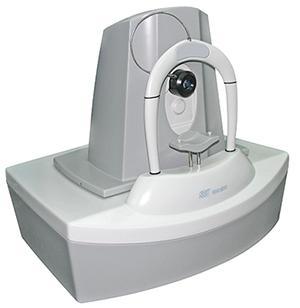
How does it work?;
Using beams of light (rather than radiation), the machine scans the retina and creates high-resolution images of its layers (interlayer imaging). This allows us to analyse both qualitatively and quantitatively the condition of the macula and optic nerve.
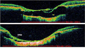
Diagnostic applications of OCT
OCT is essential for diagnosing and, more importantly, monitoring conditions such as:
Ear diseases: Age-related degeneration, edema (diabetic or postoperative), macular hole, epiretinal membrane.
Glaucoma: It offers accurate measurement of nerve fibre thickness, detecting damage long before vision is affected.
Other Diseases: Central serous chorioretinopathy, choroidal neovascularization, etc.
Frequently Asked Questions
Does the test have radiation? Is it dangerous?;
No, not at all. Although it is called a “tomography”, it does not use X-rays like a body scan. It uses simple light (low-power laser). It is perfectly safe even for pregnant women or children and can be repeated as often as needed.
Does it hurt? Is the machine touching my eye?;
The test is 100% painless and intact. You sit in front of the machine, place your chin in the slot and look at a light. The machine scans the eye from a few centimeters away without ever touching you.
Why should I do OCT and not just bottom photography?;
The bottom photo shows us the surface of the retina (like taking a picture of your skin). OCT shows us what's happening down from the surface (like seeing the layers of skin in an incision). Many conditions, such as macular edema, can only be seen clearly on OCT.
How long does it last?;
It is one of the fastest examinations in ophthalmology. The scan takes just a few seconds for each eye.
Do you need drops (mydriasis)?;
In most cases, in order to get the best possible image quality, it is preferable for the pupil to be dilated. However, modern OCT machines can often obtain satisfactory images even without drops, depending on the situation.
How does it help with glaucoma?;
In glaucoma, the damage starts insidiously from the fibres of the optic nerve. OCT can measure the thickness of these fibres. If they start to thin, it warns us about glaucoma long before your vision is lost, allowing us to start treatment early.