
Retinal Vein Occlusion: Causes, Symptoms, and Treatment
What is it?; A venous occlusion occurs when the blood flow “leaving” the eye is blocked. Usually, this is caused by compression of the vein by a hardened (atherosclerotic) artery that runs over it. The result is that the vein “bursts” from the pressure, causing:
Bleeding on the retina.
Edema in the macula (a buildup of fluid that causes blurred vision).
Ischemia (lack of oxygen), which can lead to serious complications such as neovascular glaucoma.
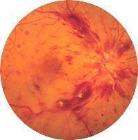
The two forms of the disease
The severity depends on the location of the blockage:
Branch Vessel Occlusion (BRVO): It blocks only a small venous branch. It affects part of the visual field and generally has a good prognosis.
Central Retinal Vein Occlusion (CRVO): The central part of the retina is blocked. The damage is extensive, vision loss is severe, and treatment is more challenging.
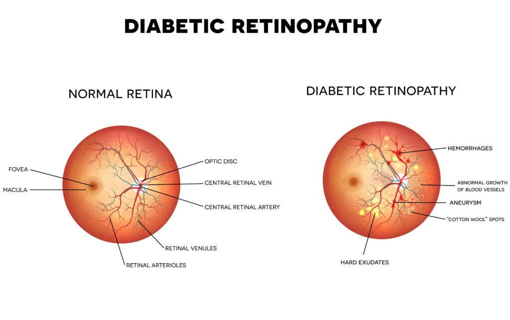
Modern Treatment (Anti-VEGF & Eylea)
The main enemy of vision in cases of blockage is the Macular Edema. Due to this disorder, VEGF factors are released, causing fluid leakage. The treatment of choice today is Intraocular Injections with factors such as aflibercept (Eylea). This medication binds to VEGF factors, “drains” the edema, and significantly improves visual acuity. In some cases, injectable steroids (cortisone) or laser treatment may also be used to treat ischemic areas.
What are the warning signs?;
Sudden blurred vision in one eye (without pain).
Blind spots or “spots” in your vision.
It's very difficult to read.
Who is most at risk?;
This condition is a “warning sign” for your overall health. It most often occurs in people with:
High Blood Pressure (the No. 1 cause).
Diabetes Mellitus.
Cardiovascular problems.
Frequently Asked Questions
Is it just as dangerous as an arterial blockage?;
No. The blockage artery It is like a stroke (interruption of blood flow) and often causes permanent blindness. The blockage vein It is similar to a blockage (overflow) and has a much better prognosis. With proper treatment, vision often returns to very satisfactory levels.
How many injections will I need?;
Treatment usually begins with one injection per month (Eylea) until the fluid has completely cleared from the macula (as seen on the OCT scan). After that, the intervals between injections are extended (every 2, 3, or even 4 months) until the condition stabilizes. Some patients may require maintenance therapy for quite some time.
Does the shot hurt?;
No. The procedure is very brief and is performed under local anesthesia. Most patients are surprised at how easy the procedure was, given how afraid they had been.
Is there a chance it could happen again?;
There is a chance of recurrence in the same eye or of the condition developing in the other eye (approximately a 10% chance). For this reason, the most important part of treatment is controlling your blood pressure and blood sugar levels, in consultation with your cardiologist or internist.
What Is Laser Treatment, and When Is It Needed?;
Laser treatment is no longer used as often to improve vision (injections are used for that). It is mainly used to “burn away” areas that are not receiving any blood supply (ischemic areas) in order to prevent serious complications such as bleeding and glaucoma.
Some days my vision is a little better, and other days it's worse. Is that normal?;
Yes. The swelling in the macula may fluctuate. However, if you notice a sudden and significant loss of vision, you should contact us immediately.