
Fluorescein angiography: The «Mapping» of the Vessels of the Eye
Fluoroangiography is an invaluable diagnostic test for evaluating the integrity of the retinal blood vessels. It allows us to identify lesions that are not visible by simple clinical examination.
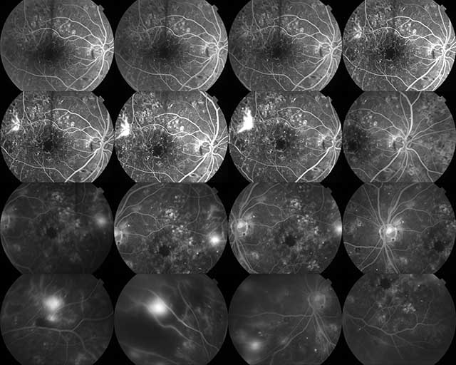
The Process
It's a quick procedure that resembles a blood draw. The doctor administers a special vegetable dye (fluorescein) into your arm. This dye acts like a “tracer”: as it circulates in your blood and passes through the eye, it illuminates the blood vessels. Our practice's special camera photographs this pathway, revealing if there are “leaks,” clogged vessels or swelling that threaten your vision.
It is the most reliable tool to decide whether you need laser treatment or intra-volvular injections.
Diagnostic Value
Fluoroangiography accurately reveals:
Fluid or blood leaks.
Swelling in the macula.
Abnormal neovessels (as in the wet form of Age Degeneration).
Areas of ischemia (poor circulation), common in diabetic patients.
The results are immediately available on the screen, allowing the doctor to plan the treatment plan on the spot.
Frequently Asked Questions
Is this dye dangerous? Is it radioactive?;
No. Fluorescein is a pharmaceutical dye and not has nothing to do with radioactive materials or iodine-containing contrast agents (such as those used in CT scans). It is generally very safe and is rapidly eliminated from the body.
I am allergic to iodine/seafood. Can I have the test?;
Yes. Fluorescein does not contain iodine. However, you should always inform the doctor of any allergies you have to medicines or foods, so that the necessary protective measures can be taken.
Does the test hurt?;
The only discomfort is the slight pinch of the needle in your hand to administer the dye (like when you have a blood test). Taking the photos is completely painless, although the camera flash is intense.
Will I have side effects afterwards?;
The most common “side effect” is that your skin and eyes may take on a light yellowish tint for a few hours. Also, your urine will have a very bright yellow-orange color for 24 hours. Don't worry, this is perfectly normal as the body eliminates the pigment. We recommend drinking plenty of water after the test.
Can I drive afterwards?;
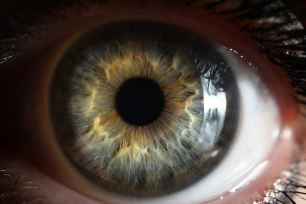
No. The test requires mydriasis (dilation of the pupil) with drops. This, combined with the bright flashes of photography, will cause blurring and photosensitivity for a few hours.
How long does the test take?;
The photography process takes about 10-15 minutes. However, you will need a total of about 30-45 minutes in the office for preparation (drops) and post-injection follow-up.