
Digital Bottom Photography (Fundus Camera)
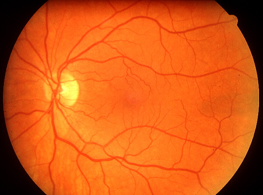
Fundus photography is the cornerstone of modern ophthalmological diagnosis. Through a special digital camera (Fundus Camera), we capture a high-resolution image of the retina, allowing for a direct and documented evaluation of the health of your eyes.
What does the test offer?;
In addition to speed and accuracy in diagnosis, the method creates a permanent digital record. This allows us to compare findings over time, accurately tracking the progression or stabilisation of a condition.
What conditions does it detect?;
The test is critical for diagnosing and monitoring conditions such as:
Age-related macular degeneration: Identification of lesions and possible neovascular membranes.
Diabetic Retinopathy: Screening for bleeding and exudates in diabetic patients.
Vascular Seals: Detection of retinal vein or artery thrombosis.
Other Diseases: Macropathies, moles and other rarer pathological conditions.
Frequently Asked Questions
Does bottom photography hurt?;
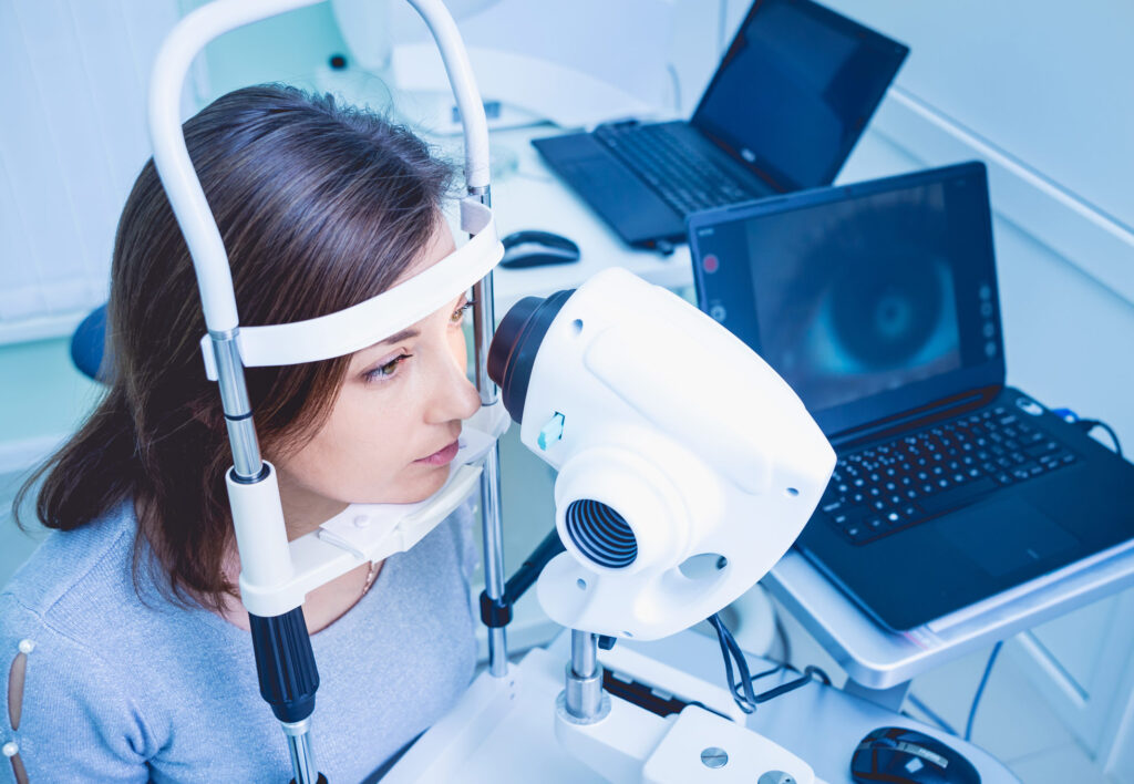
No, not at all. The process is just like when they take a normal photo of you. We don't touch your eye and there is no disturbance, other than the momentary flash of the flash.
Is flash dangerous for my eyes?;
No. The intensity of the light is perfectly controlled and safe for the eye. You may see a “spot” of light for a few seconds after the shot (as with camera flashes), but this disappears immediately.
Do you need drops (mydriasis) for the photo shoot?;
To get the best possible image quality and to see the periphery of the seabed, the use of expansion drops is usually required. However, in some cases or with special cameras, images can be taken without drops, depending on what the doctor is looking for.
How long does it last?;
Taking photos is a matter of seconds. It is usually completed in less than 1-2 minutes for both eyes.
Why is it important to keep a photo archive?;
Many conditions, such as Age-related Macular Degeneration, progress very slowly. Having the photos from your previous visit, the doctor can put them side by side on the screen and see if a lesion has grown, shrunk or remained stable. This comparison is impossible to do as accurately with clinical examination and memory alone.
Can I see the photos too?;
Of course. Immediately after downloading, the images are displayed on the computer screen. The doctor will show you exactly what he sees, explaining the condition of your eye so you can fully understand the health of your vision.