
Epoxy Membrane: Diagnosis, Statistics and Surgical Repair
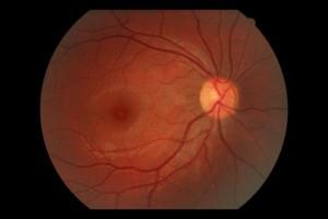
Η Epiretinal (or Epiretinal) membrane is a fibrous layer of tissue that grows on the macula. It acts as a «gelatin» that gradually hardens and shrinks, causing mechanical traction, deformation of the retina and fluid collection (edema).
Statistics & Rationale
The condition is directly related to age (idiopathic form), affecting the 3-5% of people in their 50s, a figure that rises to 20% after the 70s. It can also be caused secondarily by diabetic retinopathy, vitreous detachment, trauma or inflammation.
Diagnostic Documentation
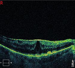
Diagnosis starts with dowsing and testing with the panel Amsler to detect metamorphopsia (crooked lines). The absolute confirmation is made through Optical Coherence Tomography (OCT).
OCT offers high-resolution, non-contact cross-sectional images, allowing the surgeon to measure the size of the membrane and the degree of edema.
Surgical Treatment (Hyalonectomy)
The treatment is exclusively surgical. This is an extremely delicate operation using insulin needle-thick instruments. The surgeon removes the membrane (peeling) with precision movements, as the area is so sensitive that it is even irritated by light.
Why is surgery considered a one-way street?;
Without surgery, vision will certainly deteriorate in 100% of cases. In contrast, surgical removal offers a total of 95% positive results:
70% of patients see a significant increase in vision (usually 2-3/10).
25% of patients achieve stabilization of vision.
Only one 5% may see a decrease in vision despite the technical success of the surgery.
Frequently Asked Questions
How urgent is the surgery for the epithelial membrane?;
The membrane develops slowly. However, the decision for surgery is made when vision begins to make daily life difficult or when OCT shows progressive deterioration. The earlier the membrane is removed, the better the functional recovery of the cells.
What are the possible complications of surgery?;
As with any fundus surgery, there is a small risk of infection (requiring immediate treatment) or retinal detachment in the first few months. Also, if you have not had cataract surgery, this procedure usually accelerates cataract formation.
Why can't my vision be corrected with glasses?;
Because the problem is anatomical (the membrane wrinkles the retina), no corrective lens can straighten the image. The solution can only be provided mechanically, by removing the membrane.
When will I be able to see the difference in my vision?;
Improvement starts after the first month, but final visual acuity is estimated at 6 months. Patience is necessary, as the retina “breathes” and heals gradually after being released from the traction.
Does the vitrectomy surgery hurt?;
No. The procedure is performed under local anaesthesia and modern small tools. Postoperatively there may be a mild discomfort (“sand” in the eye), which quickly subsides with the use of colloids.
How can I monitor the condition on my own at home?;
The best way is the table Amsler. By closing one eye, you check that the lines of the table remain straight. If you notice any new distortion or “waves”, you should contact your eye doctor immediately.