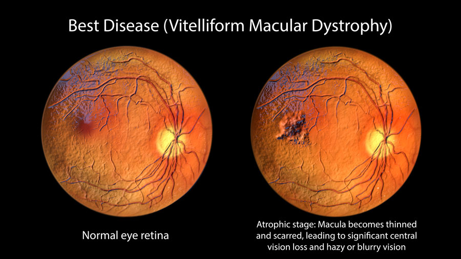
Best Disease (Vitelliform Dystrophy): from the «Crocus Egg» image to Rehabilitation
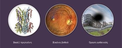
What is it?; Best's disease is an autosomal dominant disorder first described in 1905. It is caused by a dysfunction of the melancholic epithelium (RPE), which leads to an accumulation of a substance called lipofuscin under the macula.
The Clinical Pathway of the Disease
The disease progresses in stages that are often named after the appearance of the lesion:
Stage Previtelliform: Vision is excellent (10/10) and the fundus appears normal, but the diagnosis can be made with Electro-ophthalmogram (EOG).
Vitelliform stadium («Egg yolk»): A characteristic yellow round lesion appears on the macula. Vision usually remains good.
Pseudohypopyon stage: The yellow material liquefies and precipitates, creating a horizontal level.
Vitelliruptive («Omelette») stage: The damage begins to break down and vision shows the first significant bending.
Atrophic Stage: Final phase with photoreceptor loss and severe vision loss (usually after 40 years).
Diagnostic Approach
The “key” to Best disease is the EOG (Electro-ophthalmogram), which is pathological at all stages, even in carriers who have no symptoms. Η Optical Coherence Tomography (OCT) helps monitor the accumulation of material and early detection of complications such as neovascularisation.
How is vision affected?;
In childhood: Children usually have no symptoms and their vision is normal. The diagnosis is often made incidentally during a routine fundus examination.
In adult life: Vision remains functional for many years. 88% of patients retain vision capable of reading and driving in at least one eye.
After 40: Blurred vision or distortions in the lines (metamorphopsia) may occur.
What should you watch out for?;
Although there is no radical cure, regular monitoring is essential. In rare cases, new blood vessels may develop under the macula (neovascularisation), which are now effectively treated with intravaginal injections.
Frequently Asked Questions
Why is it called «egg yolk»?;
The term “vitelliform” comes from the Latin word for egg yolk. The disease is so named because in the early stages a yellow-orange mass forms in the macula that looks exactly like an egg yolk.
How will I know if my child has the disease?;
Usually the child does not complain about anything. The diagnosis is made by the ophthalmologist with fundoscopy. If there is a family history, screening should start early, as the disease is hereditary.
What is the EEG test (EOG) that the doctor requested?;
Electro-ophthalmography measures the potential of the eye during movement. In Best's disease, this test is the “gold standard” as it shows an abnormality even if the fundus appears clear. It is a painless test that confirms the diagnosis.
Will the child's daily life at school be affected?;
In most children, vision remains excellent and no special adaptations are needed. However, regular visits to the eye doctor ensure that any changes are detected early.
Is there a risk of total blindness?;
It is extremely rare. Only 4% of patients have very poor vision in the “good” eye. Peripheral vision is never affected, so the patient maintains autonomy.
How is worsening vision treated?;
If abnormal vessels (neovascularization) occur, modern ophthalmology offers the following Anti-VEGF injections, which can stabilize or improve vision. For the atrophic stage, special low vision aids are used.