Epiretinal Membrane: Cause, Diagnosis and Microsurgical Repair
What is it?; The Epiretinal (or Epiretinal) membrane is a thin layer of tissue that grows on the macula, the centre of our vision. Think of it as a «gelatin» that sits on top of the retina. As this membrane hardens and shrinks, it «wrinkles» the retina, causing distortion and blurring of vision.
What is the reason?; The most common cause is age (idiopathic form). It usually occurs after the age of 50 years, with the incidence increasing significantly (20%) after the age of 70. Other causes include diabetes, inflammation or previous trauma to the eye.

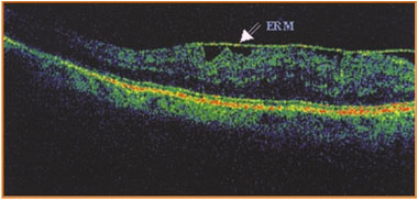
Precision Diagnosis (OCT)
The diagnosis is confirmed by Optical Coherence Tomography (OCT). This is a painless test that gives us a cross-section of the fundus, allowing us to see the thickness of the membrane and the swelling it causes, with millimetre precision.
Surgical Treatment
The treatment is exclusively surgical (Vitrectomy). In our clinic we use extremely fine instruments (thinner than an insulin needle) to remove the membrane (Peeling) and release the macula, allowing it to gradually return to its normal shape.
How does it affect vision?;
Imagine you are looking through a crumpled cellophane. This membrane exerts a pull on the macula, causing it to:
Metamorphopsia: The straight lines look crooked.
Thumb: Vision is blurred, like looking through a fog.
Diplomacy: You may see double, even with one eye closed.
The Dilemma of Intervention
Many patients are reluctant to have surgery. However, the statistics are clear:
Without Intervention: Vision will deteriorate with mathematical precision (100% probability of low vision decline).
By Intervention: You have 95% chance of improving or stabilizing vision and only 5% chance of not getting the desired result. Surgical removal is essentially a one-way street for saving vision.
Frequently Asked Questions
Is there a drug or laser to remove the film?;
No. The epiretinal membrane is a tissue (like a scar) that develops Up in the retina. It can't be dissolved by drugs or lasers. The only way to remove it is mechanically, with surgery (vitrectomy).
When will I see clearly after surgery?;
Patience is needed. Unlike cataracts where the vision clears in a few days, recovery is slow. Vision improves gradually over a period of time. 3 to 6 months. This is because the macular cells, which were “wrinkled” by the membrane, need time to straighten and function properly again.
Is the operation dangerous?;
It is a delicate operation, but with modern microsurgical tools the success rates are extremely high. Rare complications include infection or retinal detachment. Also, if you have not had cataract surgery, you are likely to develop cataracts shortly after surgery.
How much will my vision improve?;
Our objective is twofold: first, to stop the deterioration and to straighten out the “crooked lines”. Second, to gain vision. The 70% of patients gain 2-3 lines on the vision chart. Even if the vision does not become “perfect”, the image quality and reduction of distortion greatly improves daily life.
Does it hurt?;
No. The procedure is performed under local anesthesia and is painless. Postoperatively you may feel a mild discomfort (“sand” in the eye) for a few days.
How do I know if it's getting worse?;
The best way to check at home is to Amsler table (a piece of paper with squares). Close one eye and look at the center. If you see the lines getting more crooked or missing pieces, you need to call us immediately.