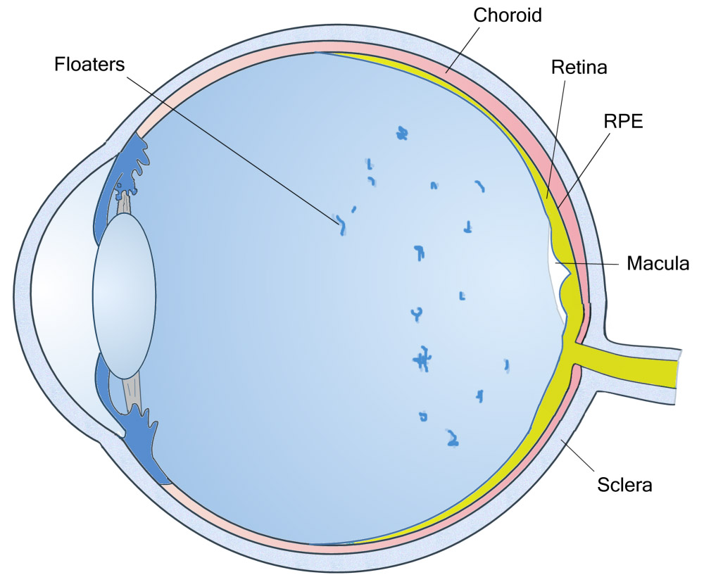
What is a vitreous?;
The vitreous is a clear gel-like substance that fills the inside of the eye, giving it structure and shape during embryonic development. Certain conditions affecting the back of the eye may require vitrectomy, namely the surgical removal of this substance. After the operation, the vitreous is replaced by normal serum, which is naturally produced by the eye.
Why is the surgery performed?;
A vitrectomy is necessary when the vitreous ceases to be clear or when it causes problems to the retina. In particular, the presence of blood (bleeding), inflammatory cells or sediment blocks the passage of light to the retina, resulting in blurred vision. The operation is also performed to remove membranes or to loosen traction forces that threaten to detach the retina from its position.

Common conditions that are treated with vitrectomy:
- Complications of diabetic retinopathy: Such as bleeding or adhesive detachment.
- Retinal detachment: To restore the tunic to its position.
- Macular hole: To close the deficit in the central area of vision.
- Epiretinal membrane: Removal of a thin membrane that “wrinkles” the retina.
- Intra-abdominal bleeding: Clearing the blood that blocks vision.
- Infections or Injuries: Treatment of serious conditions such as endophthalmitis.
- Complications of previous operations: For example, after a difficult cataract operation.
How is the process carried out?;
The ophthalmologist surgeon uses a special microscope and high-definition lenses to get a full view of the fundus. The operation is performed through three tiny incisions (a few millimetres) in the white part of the eye (sclera). The following instruments are inserted through them:
- Optical fibre: To illuminate the inside of the eye.
- Infusion line: To maintain intraocular pressure and the shape of the eye during surgery.
- Hyaloidotome: A special tool that cuts and suctions the vitreous or removes abnormal membranes.
Vitrectomy is often combined with other techniques (e.g. laser or gas/silicone injection), depending on the severity of the condition. The duration of the procedure varies depending on the complexity of the case and the general condition of the eye.
Frequently Asked Questions
Special Techniques
The vitreoretinal surgeon may use special techniques alongside the vitrectomy to treat the retina.
Your surgeon will decide if any of these are suitable for your eye.
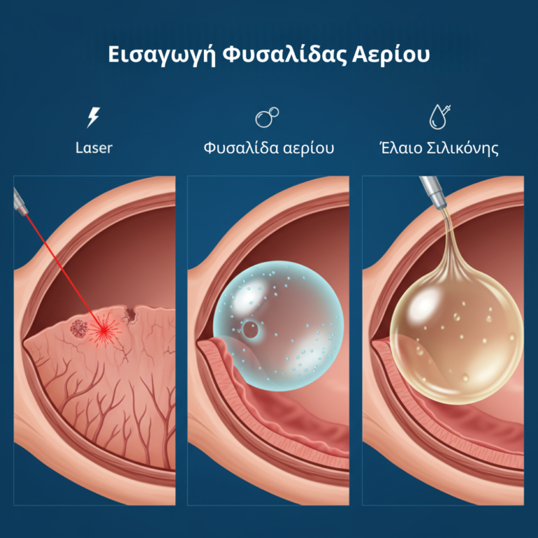
- Laser: usually used to stop bleeding in the tiny retinal vessels inside the eye.
- Gas bubble: a small gas bubble can be placed inside the eye to help close the hole in the macula or to help repair retinal detachment.
- Silicone oil: after surgery, silicone oil may be placed in your eye to keep the retina in place.
How do you feel after the surgery?;
Often there is some intolerance immediately after the operation and for a few days afterwards.
This is mainly related to the swelling on the outside of the eye and around the eyelids, which can sometimes be so large that the patient has difficulty opening the eyelids.
The abrasive sensation and occasional discomfort are to be expected.
Cold compresses are gently placed on the swollen areas to reduce the intense pain of the eye in the first few days.
Redness is common and gradually decreases with time.
Some patients may experience bruising on the outside of the eye. This is due to posterior bleeding, which has shifted under the skin and will slowly resolve on its own.
When does vision improve?;
Because vitrectomy is used to treat different types of problems and often in combination with other eye procedures, the recovery period varies depending on the case.
In some cases, such as macular hole surgery, the surgeon may place a gas bubble inside the eye, which puts mild pressure on the macula. This may require a specific head position to maintain the correct position of the bubble.
Also, mydriasis drops may be given to keep the pupil of the operated eye large, causing photosensitivity
Postoperative instructions
Because vitrectomy is used to treat different types of problems and often in combination with other eye procedures, the recovery period varies depending on the case.
In some cases, such as macular hole surgery, the surgeon may place a gas bubble inside the eye, which puts mild pressure on the macula. This may require a specific head position to maintain the correct position of the bubble.
Also, mydriasis drops may be given to keep the pupil of the operated eye large, causing photosensitivity
Can I read or watch TV after the operation?;
Yes, with conditions. Using your eyes does not cause them any harm.
Do I need to wear sunglasses?;
Since vitrectomy is usually performed alongside other techniques, postoperative instructions may vary. Some general guidelines are provided:
- Start by using antibiotic drops, which will be given to you immediately after the operation
- Avoid deep bending, weight lifting and strenuous activities.
- Follow specific instructions given for the head position (not mandatory in all cases)
However, consult your eye surgeon for specific instructions.
How long will my vision remain blurred after the surgery?;
This depends on many factors, such as the general health of your eye and the techniques used alongside the vitrectomy. Also, whether or not gas was used. Many patients notice that their vision starts to improve about three weeks after the procedure.