Displacement & Displacement of Intraocular: Modern Surgical Rehabilitation
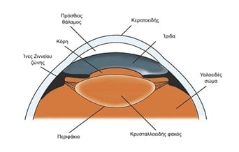
After a successful cataract surgery, the intraocular lens (the artificial lens that replaced the cloudy natural lens) must remain stable in place. However, over time, due to injury or degenerative conditions, the lens support system can weaken.
The Stages of the Shift
The process usually starts with a slight vibration of the lens (lenticular). If not treated, the lens can become completely detached from its position and fall into the vitreous cavity (the inside of the eye).
Why is the response urgent?;
Significant decrease in vision: The patient loses the ability to focus.
Risk of detachment: A lens that moves freely can come into contact with the retina, causing cracks or detachment, a condition that directly threatens vision.
Therapeutic Options & Surgical Techniques
Surgery is the only solution to restore the position of the intraocular lens. The options being considered are:
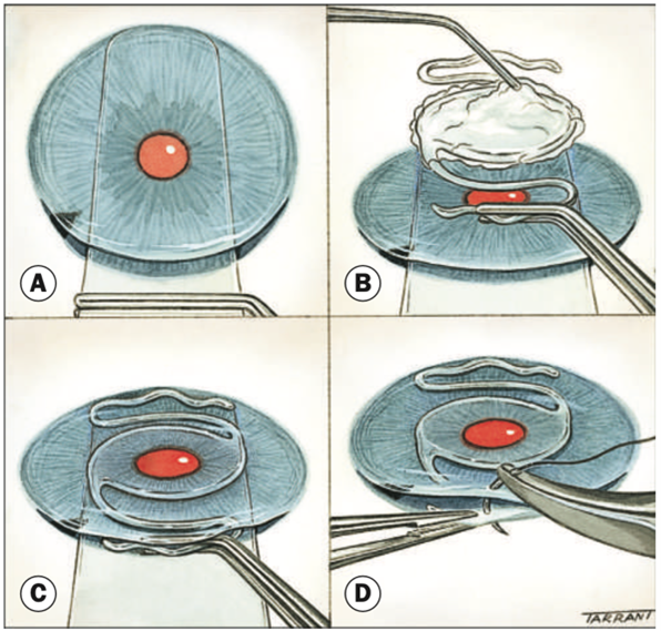
1. Repositioning of the same intraocular lens
In some cases, the existing lens is fixed back in place with special sutures. Although it is a less invasive method, it carries risks such as:
New shift or posting.
Chronic inflammation (iridocyclitis) due to contact with the iris.
Wear and tear of the material (breakage in the “legs” - haptics) or blurring of the optical body due to age.
2. Replacement with the innovative Carlevale intraocular lens
For cases where the support system is inadequate, the most modern solution worldwide is the intraocular lens Carlevale.
The advantages of the Carlevale lens
Hard support: It attaches directly to the scleral wall of the eye without the need for sutures in many cases.
Protection of the iris: It was designed to be placed in the posterior chamber without contacting the iris, avoiding inflammation.
Excellent optics: It has a large optical section that eliminates distractions and offers clear vision.
Our Experience & Scientific Work
The specialisation of the Dr. Gozzarides in Carlevale lens placement is internationally recognized.
From March 2018 to December 2019, we carried out over 60 successful placements, one of the highest numbers of cases worldwide.
Global Pioneer: We performed the first global implantation of this lens in children (aged 1 to 10 years), with results to be published in international scientific journals in 2020.
Our experience is regularly presented in Greek and international ophthalmological conferences, contributing to the development of vitreoretinal and retinal surgery.
Frequently Asked Questions
How will I know that my lens has shifted?;
The most common symptom is sudden blurred vision or the feeling that “something is moving” in your eye. Sometimes patients see the edge of the lens as a line in their field of vision.
Is the intraocular lens replacement surgery dangerous?;
It is a specialized surgery (often combined with vitrectomy), but in the hands of an experienced retinal surgeon, the success rates are extremely high and the risks are much lower than leaving the lens displaced.
Why should I choose the Carlevale lens over stitches on my old lens?;
The Carlevale lens is specially designed for eyes without support. It offers greater stability in the medium and long term, reducing the likelihood of new surgery in the future and protecting the eye from chronic inflammation.
How long is the recovery period after surgery?;
Recovery is similar to that of retinal surgery. Caution is required for about 2-3 weeks and consistency in the use of postoperative colloids.
Can the Carlevale lens be fitted to children?;
Yes, our clinic has pioneered worldwide in the application of this technique in children with excellent results, offering a permanent and safe solution in cases of congenital diseases or trauma.