Retinal artery occlusion: The «Stroke» of the Eye
What is it?; Central Retinal Artery Occlusion (CRAO) is an emergency eye condition caused when the blood flow to the retina is suddenly interrupted. Essentially, it is an ischemic episode (like a stroke) where the eye ceases to be oxygenated.
Symptoms & Clinical Picture
The characteristic symptom is the sudden, painless and complete loss of vision in one eye. On funduscopy, the retina appears white (milky) due to the swelling, while a bright red area in the centre of the macula, known as the «cherry stain».
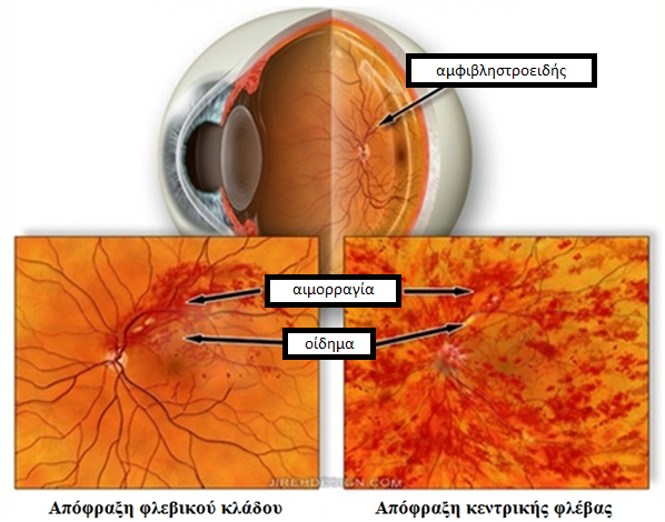
Non-productive (Substrate): This is the initial stage. The vessels are weakened and show small leaks (bleeding) or swelling in the macula, reducing vision.
Productive (Advanced): Due to poor circulation (ischemia), the eye tries to “feed” itself by creating new, poor quality blood vessels (neovascularization).
Forms of the Disease
Central Artery Obstruction: The damage is extensive and the prognosis for vision is unfortunately poor, as the retina cannot withstand the lack of oxygen for more than 1-2 hours.
Branch blockage: The piston only clogs a smaller vessel. In this case, only part of the visual field is lost and the prognosis is significantly better.
Rationale: A "bell" for your health
The blockage is usually caused by clots (blood clots or cholesterol) that travel from the heart or carotid arteries and get stuck in the eye. Therefore, this condition requires immediate investigation for cardiovascular problems to prevent a possible stroke.
Treatment: The battle with time
Immediate Response (First Hours): The aim is to move the piston. We apply techniques to reduce eye pressure (puncture, massage, drugs).
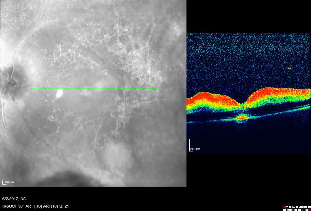
Treatment of Complications (Eylea): In cases where the ischemia causes secondary macular edema, we administer intravitreal injections with anti-angiogenic agents (such as Aflibersept/Eylea). This drug reduces the swelling and helps improve or preserve residual vision by inhibiting the VEGF factors that cause fluid leakage.
The Role of Injections (Eylea)
After the acute stage, the eye may react to the lack of oxygen by swelling (edema) in the macula. In these cases, we use the medicine Eylea (injections in the eye).
How does it help?; “It ”dries" the swelling and improves the quality of the remaining vision.
Procedure: It is painless, quick and the frequency is determined by your response (usually per month initially).
Frequently Asked Questions
Is it the same as a stroke?;
Yes, the mechanism is exactly the same. A clot or cholesterol clot leaves the heart or throat (carotid artery) and instead of going to the brain, it goes to the eye. Therefore, after the eye exam, it is necessary to see a cardiologist. Your eye ’warned“ you to protect your heart and brain.
Will I see again as before?;
At Branch blockage, vision often returns to good levels, although there may be a permanent “shadow” at some point in the visual field. At Central Obstruction, Unfortunately, the prognosis is severe and central vision rarely fully recovers unless you are one of the lucky ones who have an extra auxiliary artery (papillary artery).
Does Eylea injection hurt?;
No. The procedure is done under local anaesthesia (drops) and takes a few seconds. Most people just feel a little pressure.
How many injections will I need?;
There is no standard number. Treatment is individualized. We usually start with monthly doses and check the result with an OCT (optical coherence tomography) scan. If the swelling subsides, the injections are diluted or stopped.
Is there any way to prevent it?;
Prevention is about your general health. Controlling your cholesterol, blood pressure, blood sugar and stopping smoking are the best ways to keep your blood vessels clear and avoid blood clots in your eye or elsewhere.
What should I do if it happens again?;
If you experience sudden vision loss again (even if it's temporary and lasts for a few minutes - known as a blurring), you should go to the emergency room immediately. It's a sign that there are pebbles circulating in your bloodstream.