Retinal detachment: Symptoms, Treatment & Rehabilitation
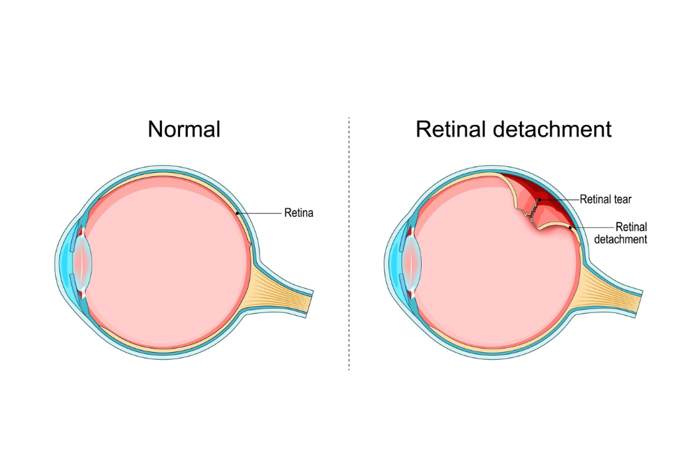
The retina is the light-sensitive membrane at the back of the eye, responsible for converting images into a signal to the brain. Its detachment is an emergency condition that requires immediate treatment to save vision.
How is it created?;
It usually starts from a crack. The vitreous (the gel that fills the eye) liquefies with age and detaches from the walls. If at any point it is “hooked” onto the retina, it can pull and tear it. Through this tear (crack), fluid passes through which “pulls” the retina out of place, just as wallpaper comes off the wall.
The 3 Warning Symptoms (SOS)
If you notice any of the following, you should see an eye doctor immediately:
Flashes: Like camera flashes or lightning flashes, usually in the periphery of vision.
Bugs: Sudden appearance of many black spots or “spiders” that move.
Curtain: A shadow that starts from the side and begins to cover the picture (as if a curtain is falling).
Therapeutic Treatment
If you are going to have surgery for cataracts, this is the examination that will determine the lens we put in your eye.
- At the crack stage (without detachment): If we catch the damage early, treatment is easily done in the clinic with Laser Photocoagulation. The laser “glues” the area around the crack, preventing it from expanding.
- At the detachment stage: The treatment is exclusively surgical. Today, thanks to modern techniques (Hyalonectomy), the success rate exceeds the 90% with a single operation.
The Procedure (Hyalonectomy)
This is a “microsurgical” technique (usually without stitches).
We remove the vitreous and the tractions causing the damage.
We return the retina to its position (Laser or Cryocoagulation).
We fill the eye with special gas ή silicone oil to keep the retina attached until it heals.
Frequently Asked Questions
Does the operation hurt?;
No. The procedure is usually performed under local anaesthesia and intoxication, so the patient does not feel any pain. Postoperatively there may be a slight foreign body (“garbage”) sensation, which is easily treated with painkillers and eye drops.
What is a "gas bubble" and when does it go away?;
At the end of the surgery, we often fill the eye with a special gas. This acts like an “internal splint”, pushing the retina to stick. The gas is absorbed by the body on its own over a period of 2 to 4 weeks and does not need to be removed.
Why do I have to look down or lie down after surgery?;
If you have been gassed, the position of the head is critical. The gas always rises upwards (like a balloon). To press the right spot on the bottom, you must bring your head to the appropriate position indicated by the doctor. Compliance with this greatly increases the success of the procedure.
When will my vision return?;
Recovery is not immediate. As long as the gas bubble is present, you will see very blurry (as if in water). As the gas goes away, you will see a “line” go down and your vision will clear from top to bottom. Final improvement may take 3 to 6 months.
Can I travel by plane?;
ATTENTION: As long as you have gas in the eye, is strictly prohibited travelling by plane or climbing a high altitude (mountain). The change in atmospheric pressure causes the gas in the eye to expand, with the risk of serious damage and pain. You will only be able to fly when the doctor confirms that the gas has been fully absorbed.
If you put silicone in me instead of gas, what difference does it make?;
In more complicated cases, we use silicone oil. The difference is that the silicone does not go away on its own. It offers stability for a long time, but we will need a second, shorter procedure after a few months to remove it. With silicone you can travel by plane.